
Imagine truly understanding your dental health, not just being told about it. For generations, a trip to the dentist meant a lot of trust in what you were told, perhaps glimpsing an X-ray, but rarely getting a clear, up-close look at your own teeth. Today, that’s all changing, and it’s making dental care more transparent, educational, and effective than ever before. In the world of modern dentistry, X-rays are undeniably crucial – they help us see what’s happening inside your teeth and under your gums, like a blueprint of your oral foundation.
But what if you could also take a guided tour of the surface of every tooth, magnified a hundred times, in real-time?
That’s the magic of advanced intraoral cameras, and they’re revolutionizing how we detect issues, educate patients, and craft personalized treatment plans.
Your Mouth, Magnified: How Advanced Intraoral Cameras Work
Think of an intraoral camera as a tiny, pen-sized wonder-tool that brings a super-magnified, high-definition view of your mouth right to a screen in front of you. It’s not magic, it’s brilliant engineering!
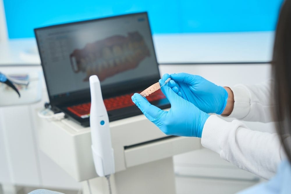
These miniature cameras are equipped with their own LED light source, which brightly illuminates even the darkest corners of your mouth.
As the dentist gently glides the camera wand over your teeth and gums, it captures incredibly detailed, crisp images and video.
These visuals are then displayed instantly on a monitor, allowing both you and your dental team to see precisely what’s happening.
This technology offers a fundamental advantage: magnification.
While a traditional mirror and probe allow your dentist to see a lot, an intraoral camera can magnify details up to 100 times, revealing minuscule cracks, subtle discoloration, or early signs of gum inflammation that might otherwise be missed by the naked eye.
This level of clarity is the first step in a new era of proactive oral health.
More Than Meets the Eye: Why Intraoral Cameras Are a Game-Changer
While standard intraoral cameras provide fantastic visual clarity, advanced models go much further, offering sophisticated diagnostic modes that uncover problems long before they become serious.
The Power of Unprecedented Clarity: Seeing 100x Closer
With crystal-clear, high-resolution images, we can spot incredibly fine details that are easy to miss. This includes:
- Micro-cracks: Tiny fractures in the enamel that can grow into bigger problems if not addressed.
- Worn or failing fillings: Old fillings that are chipping, leaking, or nearing the end of their lifespan.
- Early gum inflammation: Subtle redness or puffiness around gum lines, indicating the very beginning of gingivitis.
- Plaque and tartar buildup: Areas you might be missing during brushing, clearly visible and explainable.
Seeing these magnified issues can be an “aha!” moment for many patients, transforming abstract concepts into tangible realities.
It helps you understand why a certain recommendation is being made, making you an active participant in your care.
Revealing Hidden Threats: Advanced Diagnostic Modes
Some of the most powerful advancements in intraoral camera technology come from specialized lighting techniques that allow us to detect issues before they’re visible to the naked eye or even show up clearly on traditional X-rays.

- Fluorescence Technology (like LIF or “Cario” mode): This isn’t just about taking a picture; it’s about seeing biological activity. Certain advanced intraoral cameras use specific wavelengths of light that make healthy tooth structure fluoresce one color, while bacterial byproducts from early decay or plaque fluoresce a different color (often red or orange). This means we can literally “see” early decay glowing under a special light, pinpointing demineralization before it becomes a full-blown cavity. Research from the National Institutes of Health (NIH) highlights how intraoral cameras significantly increase the detection of occlusal caries (cavities on the chewing surfaces) in studies, leading to more correct treatment decisions.
Transillumination: Another clever trick! This technique involves shining a powerful, focused light through the tooth. Healthy tooth structure allows light to pass through evenly. However, a hidden crack or an interproximal cavity (a cavity between teeth) will block or scatter the light, appearing as a dark shadow or line. This is incredibly effective for identifying problems that might be missed by other methods.
Your Oral Health Story, Documented
Beyond immediate diagnosis, intraoral cameras create a visual record of your oral health journey.
These images can be stored securely, allowing your dental team to:
- Track changes over time: Monitor suspicious spots to see if they’re progressing or staying stable.
- Document before and after: Show the incredible transformation after procedures like fillings, crowns, or [cosmetic dentistry treatments]([Internal Link Placeholder: Cosmetic Dentistry/Cosmetic Dentistry]).
- Communicate effectively: Share images with specialists if a referral is needed, ensuring everyone is on the same page.
Intraoral Camera vs. X-ray: Allies in Your Oral Health Journey
A common misconception is that intraoral cameras replace X-rays.
This couldn’t be further from the truth! They are highly complementary tools, each with unique strengths.
- X-rays are like looking at the foundation and internal structure of a house. They use radiation to show us what’s happening inside your teeth (like deep cavities, root issues), and under your gums (like bone levels, impacted teeth, or infections). They are indispensable for a complete diagnostic picture.
- Intraoral cameras are like walking through that house with a super-bright flashlight and magnifying glass, meticulously checking every surface and corner. They excel at revealing surface-level issues, gum health, and overall oral hygiene. Crucially, they use no radiation, making them a fantastic, safe option for frequent checks, especially for surface concerns or for patient groups like pregnant women and children.
By combining the insights from both X-rays and intraoral cameras, your dental team gets the most comprehensive view possible of your oral health, allowing for the most accurate diagnoses and effective treatment plans.
The Co-Diagnosis Advantage: Becoming an Active Participant in Your Care
One of the most profound benefits of advanced intraoral cameras is the way they empower you, the patient.
When you can see what your dentist sees, a new level of understanding and partnership emerges.
Studies, like those published by the NIH, suggest a positive impact on patient compliance with dental care when intraoral cameras are used.
Seeing an issue directly on a screen in real-time, magnified and clear, makes it undeniable. It helps to:
- Demystify diagnoses: No more guessing games about what a “cavity” or “gum inflammation” truly looks like. You’re shown the evidence.
- Build trust and transparency: When your dentist points to an issue on the screen and explains it, you become an active participant in your diagnosis, fostering a sense of partnership and trust. This transparency helps build confidence and enables people to take an active role in their health.
- Inform better decisions: With a clear visual understanding, you’re better equipped to discuss treatment options, ask questions, and make informed choices about your oral health journey. Whether you’re considering a filling, [Invisalign for a straighter smile]([Internal Link Placeholder: Invisalign/Invisalign treatments]), or veneers, seeing the starting point clearly helps you envision the outcome.
This “co-diagnosis” approach ensures that your treatment plan isn’t just something done to you, but rather a collaborative decision made with you.
Beyond the Camera: What Advanced Imaging Means for You
The impact of advanced intraoral cameras extends far beyond early detection. They represent a fundamental shift towards more preventative, precise, and patient-centered dentistry.
- Preventing Bigger Problems: Catching a tiny crack or early demineralization with an intraoral camera means we can often intervene with minimally invasive treatments, like a small filling or a fluoride application, potentially preventing the need for a larger, more complex, and more costly procedure like a crown or even a root canal later on.
- Long-Term Monitoring: These detailed images serve as invaluable benchmarks, allowing us to accurately track the progression of non-urgent issues, monitor healing, and confirm the stability of your oral health over years. This long-term perspective is a cornerstone of true [preventative dental care]([Internal Link Placeholder: Preventative Dental Care/Preventative Dental Care]).
- Holistic Digital Dentistry: Intraoral cameras are an integral part of a modern, holistic digital dentistry ecosystem. Their images can integrate with digital X-rays, 3D scanners, and even CAD/CAM technology for same-day crowns, creating a seamless and highly efficient patient experience.
Your Questions Answered: Intraoral Cameras FAQ
Q1: Is an intraoral camera safe to use? Does it expose me to radiation?
Absolutely! Intraoral cameras are completely safe. Unlike X-rays, they do not use any ionizing radiation. They simply use visible light, much like a tiny flashlight, to illuminate your mouth and capture images.
Q2: Will the intraoral camera procedure be uncomfortable or painful?
Not at all. The camera wand is small and designed to be non-invasive. Your dentist will gently guide it around your mouth to capture images, and most patients find the experience entirely comfortable.
Q3: Do intraoral cameras replace the need for traditional X-rays?
No, they are complementary tools. X-rays provide crucial information about the internal structure of your teeth and bone, which the intraoral camera cannot see. The camera excels at examining the surface of your teeth and soft tissues. Together, they give your dentist a complete picture of your oral health.
Q4: Can an intraoral camera really detect all types of cavities?
Intraoral cameras, especially those with advanced diagnostic modes like fluorescence and transillumination, are excellent for detecting early cavities on tooth surfaces and between teeth. However, deep cavities that extend into the dentin or are hidden beneath existing fillings might still require an X-ray for full assessment.
Q5: How does seeing the images on the screen help my dentist plan my treatment?
Seeing magnified, clear images helps your dentist precisely identify issues and explain them to you. This shared understanding allows for “co-diagnosis,” where you and your dentist can discuss treatment options more effectively, leading to more informed decisions and personalized care tailored to your specific needs.
Ready to See Your Oral Health in a New Light?
The advanced intraoral camera is more than just a piece of technology; it’s a window into a clearer, more informed, and proactive approach to your oral health. It empowers you to understand your smile better, participate actively in your care, and ultimately, make the best decisions for your well-being.
At Radiance Artistic Dental & Wellness in Boulder, Colorado, we believe in ethical dentistry that builds trust and fosters understanding. We integrate advanced tools like intraoral cameras into our holistic approach, ensuring you receive the most comprehensive, transparent, and personalized care possible.
If you’re curious to experience modern dentistry that puts you in the driver’s seat of your oral health journey, we invite you to learn more.


